Dental radiographs, usually referred to as dental x-rays, are a crucial tool in contemporary dentistry. They give dentists important information about dental health and help them in diagnosis and treatment planning. In this blog post, we’ll examine the many dental X-rays kinds and how each one contributes to maintaining a beautiful smile.
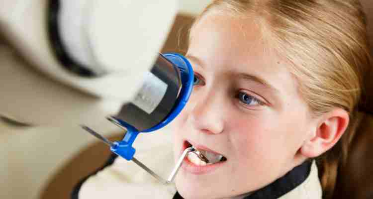
X-rays of the bitewings: One of the most frequent dental X-rays taken during a normal examination is a bitewing X-ray. The reason they are called “bitewing” X-rays is that the patient bites down on a tiny piece of paper or a piece of plastic that keeps the X-ray film in place, enabling the dentist to take a single image of the upper and lower back teeth. In particular, bitewing X-rays are used to:
1. Look for dental caries or cavities between teeth.
2. Evaluate how well dental crowns or fillings fit.
3. Tracking the development of gum diseases.
Periapical X-rays: As the name implies, a particular location is the focus of the X-ray. The entire tooth is visible in these X-rays down to the tip of the root and the surrounding bone. Using periapical radiography is necessary for:
1. Recognizing infections or abscesses at the root of the tooth.
2. Analysis of the bone health and state of the root.
3. Examining possible issues with impacted teeth.
Panoramic X-rays: These images provide a comprehensive view of the entire mouth. They are made by placing the patient’s head inside a revolving apparatus that captures a sizable image. Invaluable uses for panoramic X-rays include:
1. Verifying the growth and placement of every tooth, including wisdom teeth.
2. Identifying jaw problems such as temporomandibular joint (TMJ) abnormalities.
3. Making preparations for oral surgery or orthodontic therapy.
Cone Beam Computed Tomography (CBCT): A more sophisticated type of dental imaging, CBCT produces three-dimensional (3D) images of the teeth and their surroundings. Uses for CBCT scans include:
1. Dental implant planning.
2. Assessment of difficult dental problems, such as malignancies or impacted teeth.
3. A thorough evaluation of bone volume and density for oral surgery.
Occlusal X-rays: It takes a wide-angle picture of the mouth’s floor, usually concentrating on the bite. These X-rays can help with:
1. Identifying more teeth that may not have fully erupted.
2. Detecting fractures or other problems with the mouth’s roof or floor.
3. Examining young toddlers whose bites are still establishing the position of their teeth.
Extraoral X-rays: These X-rays are taken from outside the mouth and are primarily used to visualize the skull and jaw. Common types include:
1. Cephalometric X-rays are used in orthodontics to evaluate the growth of the face and teeth.
2. Sialography: This procedure involves injecting a contrast dye to make the salivary glands visible.
3. Tomograms: Offer a thorough image of a particular layer of the jawbone or mouth.